
Being pregnant is a wonderful feeling but it has its phases of apprehension. You probably have a whole questionnaire ready about the growth of baby, symptoms of pregnancy, dos and don’ts of pregnancy and whatnot.
One of the commonest aids used by health providers over the world to visualize pregnancy and the growth and development of the fetus is ultrasonography, commonly referred to as ultrasound.
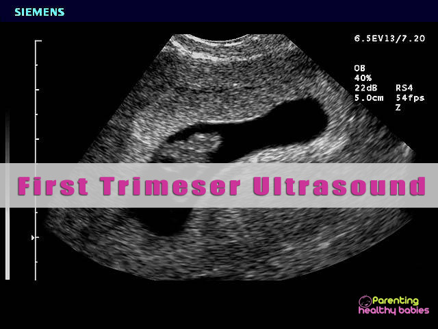
Getting an ultrasound done for the first time can be a flustering experience. Knowing what to expect and being prepared in advance can make it an amazing and delightful experience.
First Trimester Ultrasound Scans
About Ultrasound:
Ultrasound is a simple painless procedure which uses sound waves bounced back from internal organs and fetus to envision them.
It consists of a transducer (a small probe like object) and a monitor. The health care provider will spread a cold gel on the abdomen and place the transducer on the area. The transducer emits sound waves into the abdomen which travel through liquid to the fetus. The fetus reflects these waves back to the transducer. These reflected waves are converted to pictures or video which can be seen on the monitor or printed.
When to Have an Ultrasound?
Soon after you get a positive pregnancy test and visit your doctor, the doctor will schedule an ultrasound scan.
Usually two ultrasound scans are done in the first trimester:
- Dating and viability scan: between 6th to 9th
- Morphology or NT (nuchal translucency) scan: between 11th to 13th
Types of Scans:
- The ultrasound scans done before the 10th week are usually trans-vaginal scans. The fetus is too small to be seen through the abdomen, so it is seen through the vagina. Trans-vaginal scan can also help us to see internal organs of the mother.
- The scans after the 10th week are generally abdominal scans. They are done by placing the transducer on the lower portion of the abdomen.
Preparing for an Ultrasound Scan:
- In an ultrasound scan, the lower part of the body needs to be exposed. It is advised to wear loose, comfortable two piece clothing.
- In the first trimester, the fetus is small. To see it properly, the sound waves need to travel through a liquid medium. It is imperative to ask your health care provider if you need to keep the bladder full before the scan.
- Generally trans-vaginal scans are done on an empty bladder.
- Abdominal scans, on the other hand, require a full bladder. Some providers suggest a half full bladder. For this, empty your bladder two hours before the scan. Over the next hour, drink about 1 liter water. Do not go to the toilet unless instructed.
Dating and Viability Scan: Importance:
Dating and viability scan is the ultrasound done between the 6th and the 9th week from LMP. The importance of this test is:
- To check if the location of the fetus is correct. Sometimes, fetus may be implanted in the fallopian tube instead of the uterus. This is a dangerous condition which needs to be detected and treated accordingly. Don’t worry; it occurs in just 1% of pregnancies.
- To check the heart beat and development of the fetus.
- To determine an accurate due date especially in women with irregular menstrual cycles.
- Number of babies!
The scan should not be skipped in some conditions like:
- If you have irregular cycles
- If your doctor suspects ectopic pregnancy or you have a history of ectopic pregnancy
- If you have had a miscarriage
- If you have vaginal bleeding
Multiple scans may be scheduled by the doctor in the following conditions:
- Persistent vaginal bleeding
- Multiple pregnancy
- If the mother is over 35 years of age and this is her first pregnancy
- If the mother has fibroids, ovarian cyst etc.
Nuchal Translecency Scan:
NT test is a two part test done in the 11th to 13th week of pregnancy. The first part consists of an ultrasound scan and the second part is a simple blood test where various enzymes, hormones, protein levels and other markers are analyzed.
In the ultrasound scan part, various things which are examined are:
- Position of placenta
- The thickness of the folds of the back of neck (nuchal translucency thickness)
- Presence or absence of nasal bone
- Problems with fetal spine and limbs
- Fetal abdominal defects
- Blood circulation to mother’s uterus.
These are the markers of Down’s syndrome, trisomy 18, developmental spinal and heart defects.
Ultrasound Scans: Week by Week
- Mid 4th week: A small circle may be seen in the center of the sonogram. That is the gestational sac. It appears black because it is filled with fluid.
- 5th week: A white circle can be seen inside the gestational sac. This is the yolk sac. A transvaginal scan is required to see the baby. It is just a few millimeters long. Almost the size of a bean!
- 6th week: The fetus looks bigger. It is 5-9 mm long. The crown-rump length (CRL) of the fetus is measured now. The baby’s heartbeat can be detected.
From 6th week to 9th week the baby grows at a constant rate of 1 mm per day. CRL is the primary marker used to calculate the due date until the 13th week. After 13th week, the due date is calculated by bi-parietal diameter (BPD) of the head of the baby
- 7th week: The baby is seen clearly. The heartbeat can be measured to be around 120 per minute.
- 8th to 10th week: The baby is now seen by the abdominal ultrasound. It is now 1-2 cm in length
- 11th to 12th week: The baby is clearly recognizable with a well-defined body, head, arms and legs. It may be seen moving around in the gestational sac.
Read more: 3D/4D Ultrasound During Pregnancy
Safety of Ultrasound:
Ultrasound does not contain any radiations unlike X-rays and is absolutely safe for both the fetus and the parent when done by a trained professional. Doctors advise against the 3D photo-shoot ultrasound carried out in photo booths because of untrained staff.
Happy parenting!
Hope this article was of help for all our parents!! Please share your comments/queries/tips with us and help us create a world full of Happy and Healthy Babies!!


